Smile Gallery

Treatment journeys & surgical results

Follow selected cases from the first visit to the final result — what the problem was, what was done, and how it turned out.

Treatment Journeys

From first visit to final result

Each case below shows the same patient at four stages — before, during, after and at follow-up.

Case 01 · Trauma Diagnosis
The case

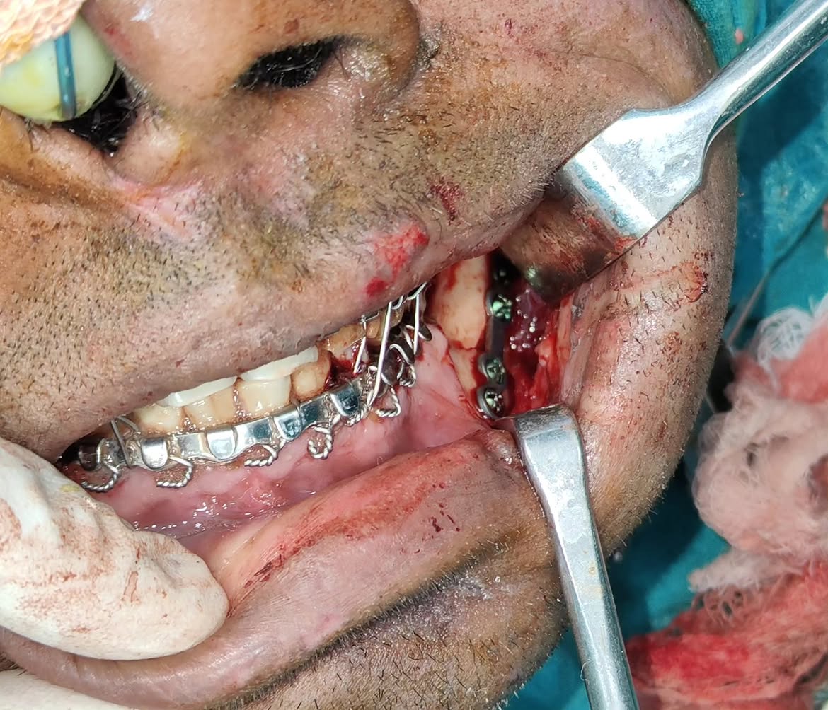
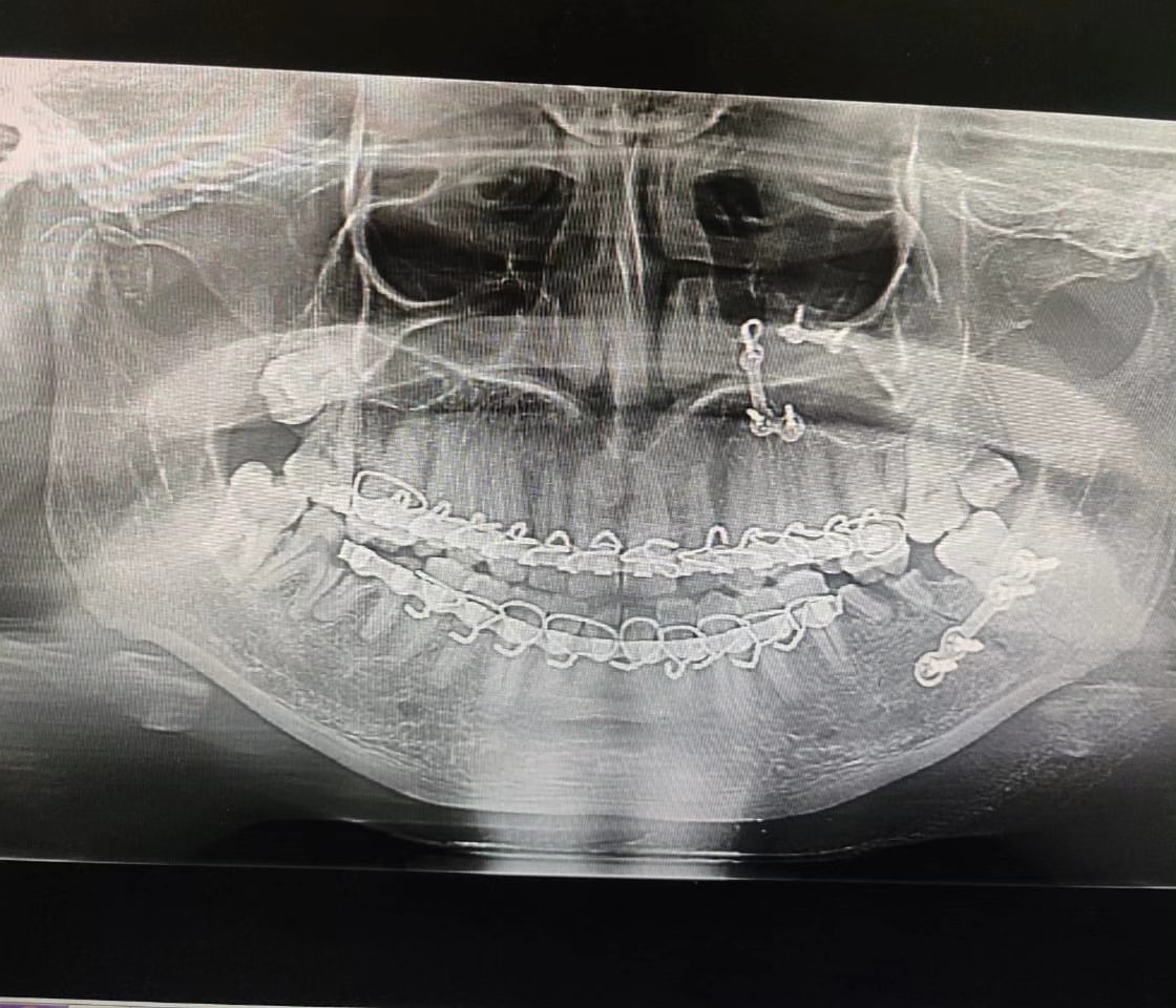
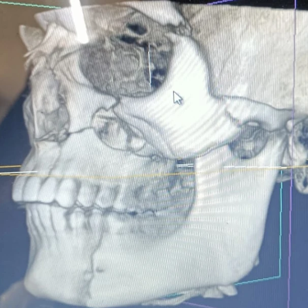
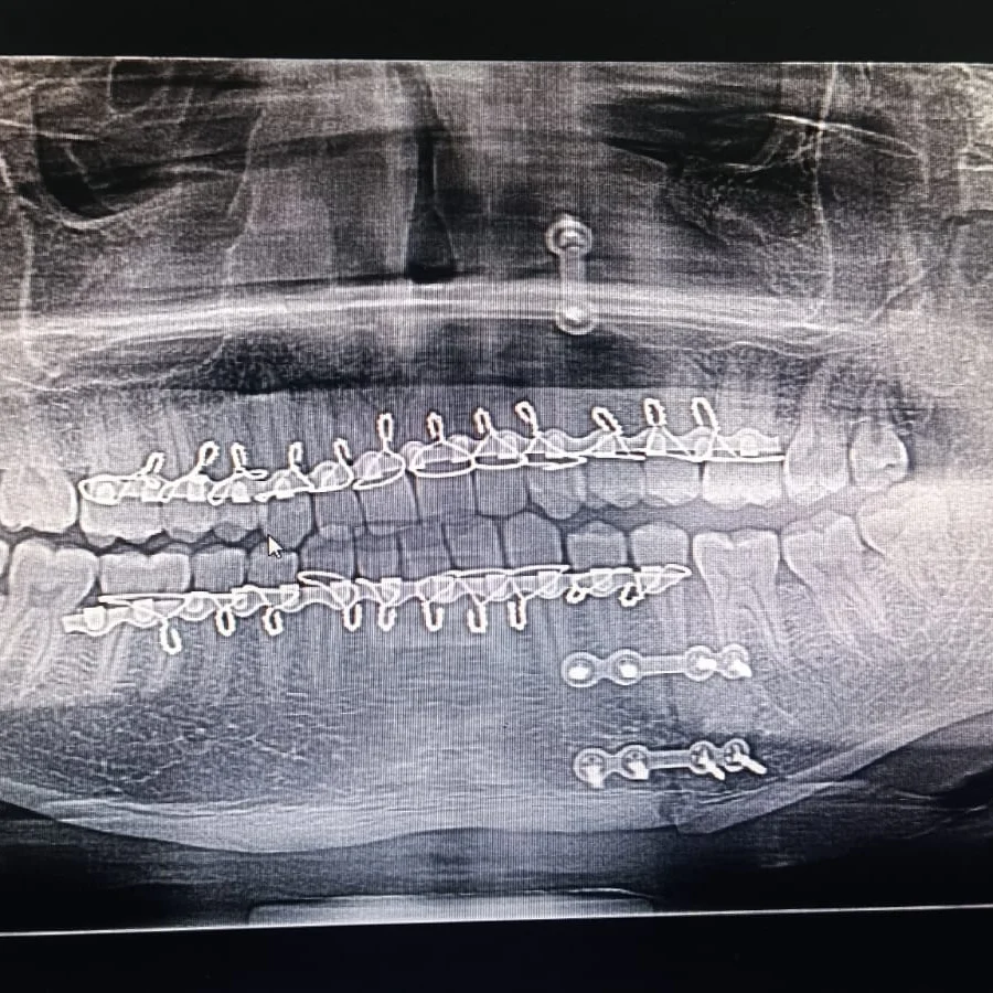
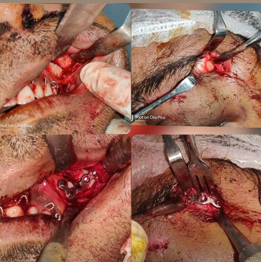
Case of trauma Diagnosis -fracture left body of mandible, Fracture left maxillary pyriform rim and buttress, Fracture left infraorbital rim.

The treatment

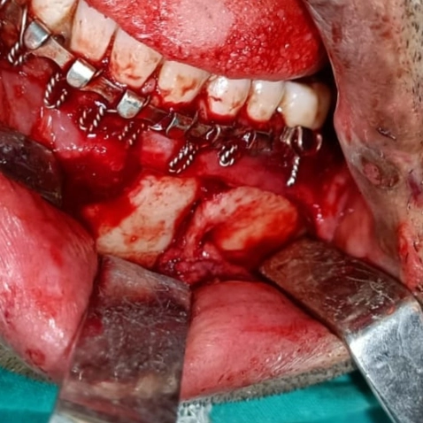
Treatment done-open reduction and internal fixation under general anesthesia Incision taken-infraorbital incision givem to expose left infraorbital rim, Vestibular incision given to expose left maxillary pyriform rim, Modified Ward's incision given to expose left body region.

The outcome

IMF done and occlusion achieved. Fixation done using miniplates and miniscrews. Case done
Case 1: jaw cyst before treatment — Dr. Mahesh Pund
Before
Case 1: jaw cyst during surgery — Dr. Mahesh Pund
During
Case 1: jaw cyst immediately after surgery — Dr. Mahesh Pund
After
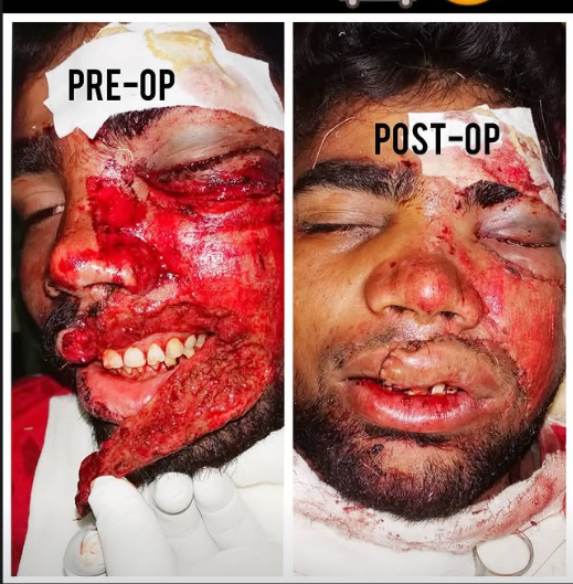
Case 02 · Facial Trauma Surgery

Jaw fracture fixation — restoring bite and facial alignment

The case

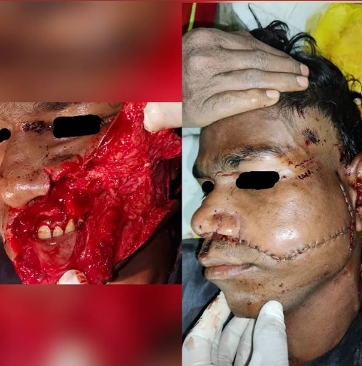
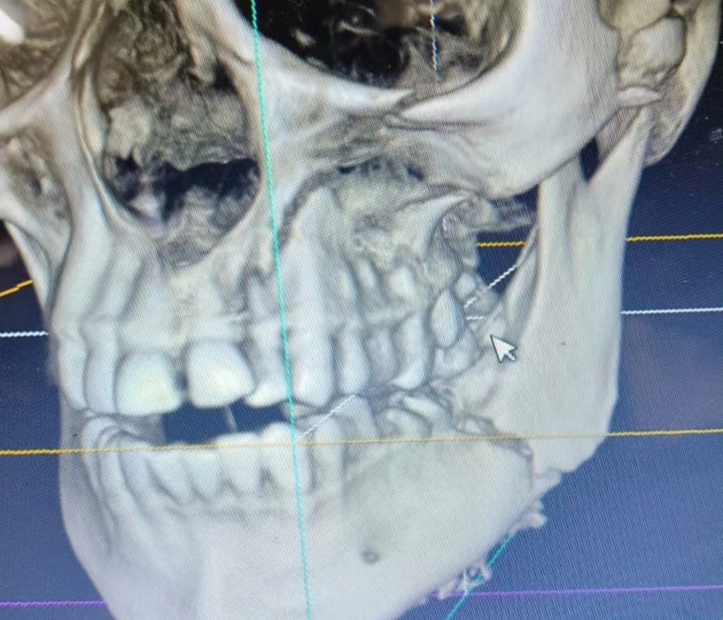
The patient presented with facial trauma and was diagnosed with a left-sided Le Fort II fracture along with a left parasymphysis fracture.

The treatment

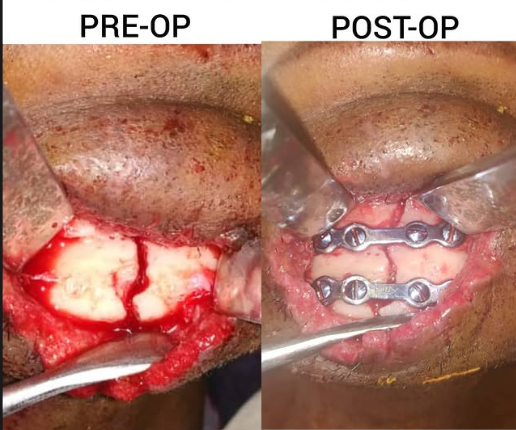
Open reduction and intermaxillary fixation were performed. Lateral brow and circumvestibular incisions were used to access the fracture sites, followed by fixation using miniplates and miniscrews.

The outcome

The fractures were stabilized with 2 mm miniplates and miniscrews, restoring proper alignment and providing stable fixation for healing.

Case 2: jaw fracture before treatment — Dr. Mahesh Pund
Before
Case 2: jaw fracture during surgery — Dr. Mahesh Pund
During
Case 2: jaw fracture immediately after surgery — Dr. Mahesh Pund
After
Case 03 · Dental Implant

Implant with bone grafting — rebuilding bone for a lasting result

The case

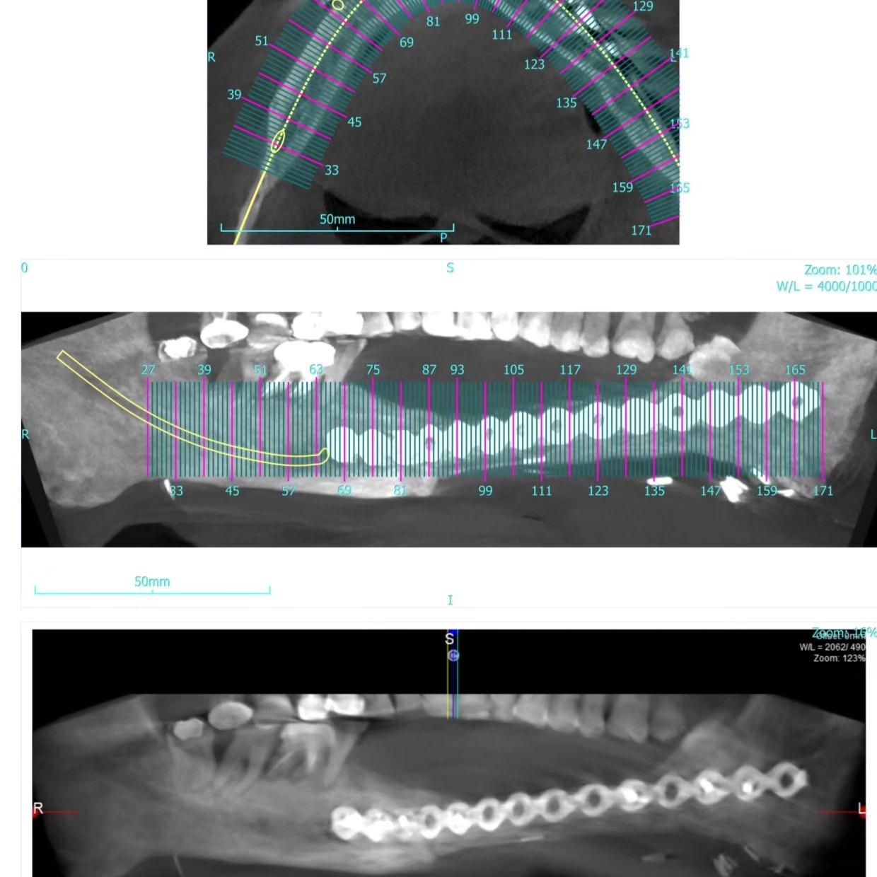
Patient had missing teeth following reconstruction with a free fibular graft after carcinoma surgery 7 years ago. CBCT was used to assess the grafted bone and plan implant placement.

The treatment

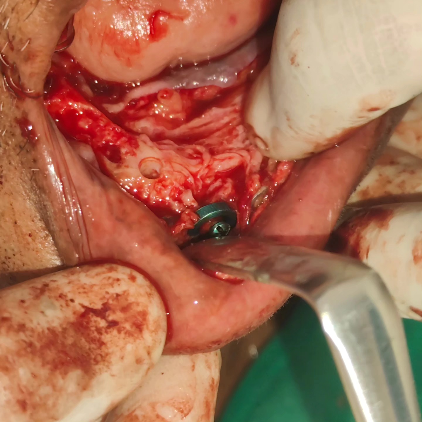
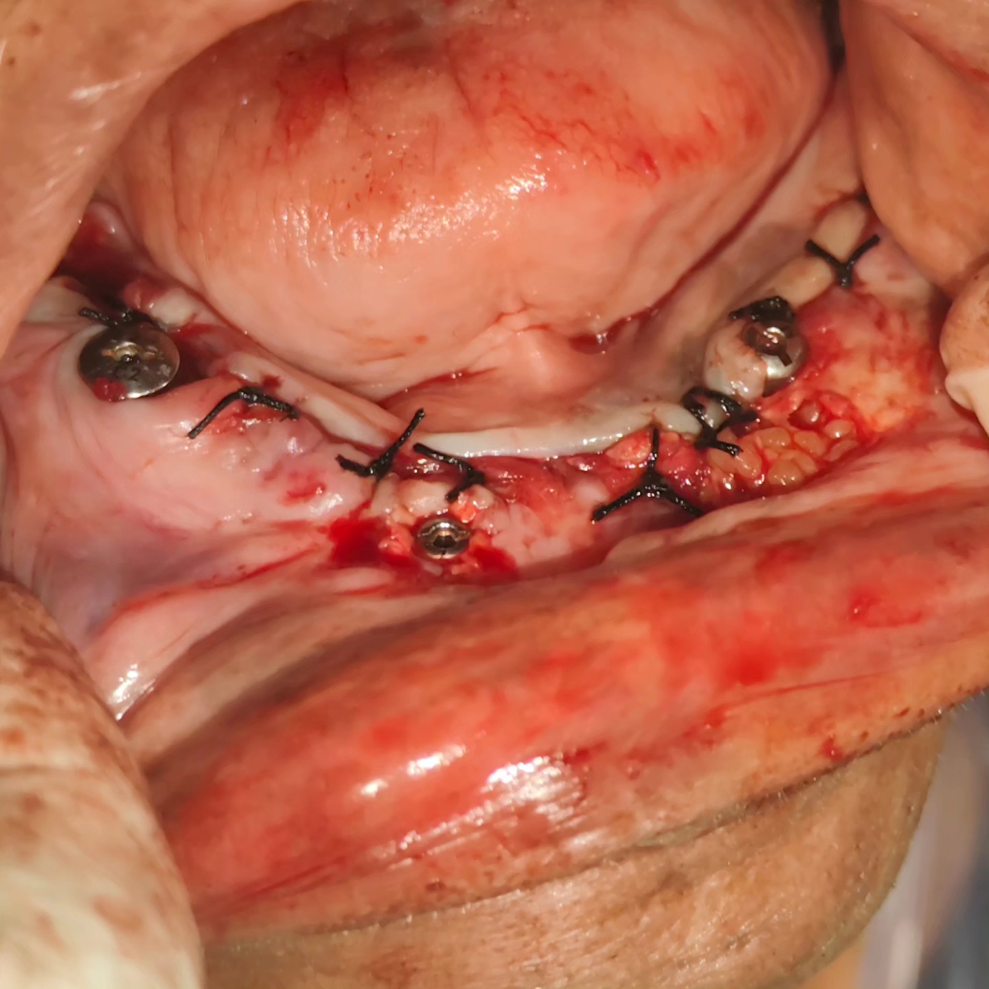
Three implants were placed in the reconstructed fibular graft: two 4.5 × 8.5 mm implants in regions 34 and 45, and one 4 × 10 mm implant in the indicated region.

The outcome

The implants were allowed to integrate with the grafted bone before final restoration, providing a stable foundation for replacement teeth with improved chewing function and appearance.

Case 3: implant with bone grafting before treatment — Dr. Mahesh Pund
Before
Case 3: implant with bone grafting during surgery — Dr. Mahesh Pund
During
Case 3: implant with bone grafting immediately after surgery — Dr. Mahesh Pund
After
Selected Cases

More treatments at a glance

One image per treatment — a quick look at other procedures performed.

Impacted Wisdom Tooth Removal — Dr. Mahesh Pund
Dental Implant — Dr. Mahesh Pund
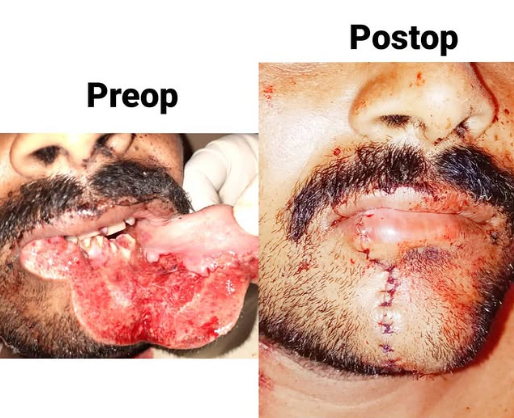
Oral Lesion Removal — Dr. Mahesh Pund
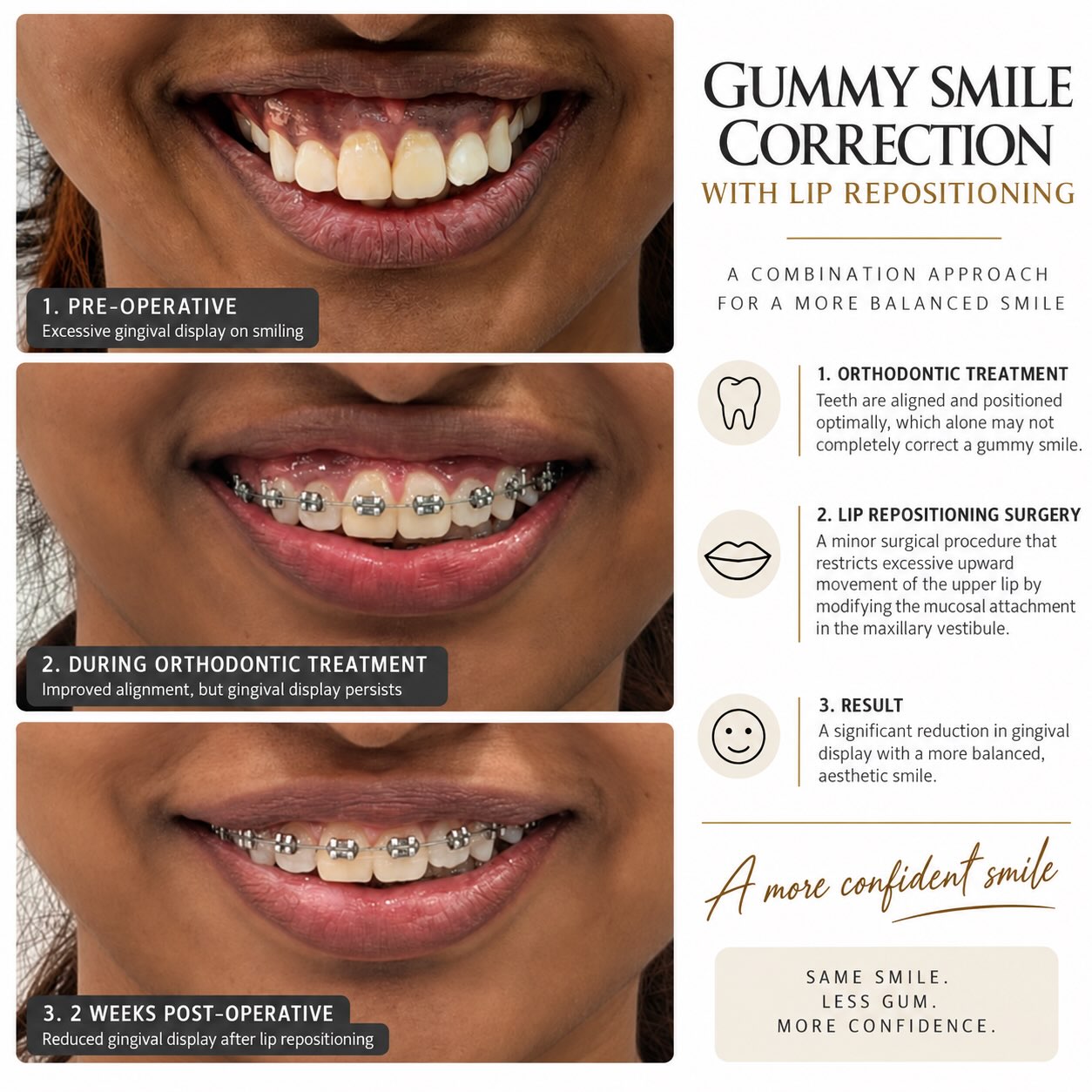
Gummy Smile Correction — Dr. Mahesh Pund
Facial Fracture Fixation — Dr. Mahesh Pund

Considering a procedure?

Discuss your case directly with Dr. Mahesh Pund.